Manuka Honey

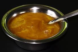
Background Honey has been long utilised as a topical antimicrobial treatment for wound infection, with licensed medical products becoming available in the 1990’s. Whilst often still used as a last line treatment when other therapies have failed, manuka honey is broad spectrum with potent bactericidal activity. Some of the bacterial cellular targets for manuka honey are now known and more continue to be identified. For Gram positive organisms the primary mode of action is to disrupt the cell cycle leading to aberrant cell division and weakening of the cell wall; this is combined with an up-regulation of the general stress response [1,2]. In Gram negative organisms, particularly Pseudomonas aeruginosa, the major targets appear to be integral membrane proteins such as OprF which ordinarily stabilise the cell leaflet and without which, bextensive membrane disruption and eventual lysis occurs [3].
Essentially, studies have demonstrated that with short- and long-term training experiments, no resistance to manuka honey treatment emerges [4,5]. This is of particular significance in an age where antimicrobial resistance outstrips the rate at which new antimicrobial treatments are discovered and where there is real concern that species resistant to all known antibiotics could become rife. The problem of resistance can be exacerbated, for example when microorganisms grow as a biofilm. Such organisms are afforded intrinsic protection in the form of an extra-polysaccharide layer which restricts the diffusion of antimicrobials meaning that therapeutic doses do not reach all bacteria within the biofilm, causing infection to recur [6,7]. Therefore the biofilm mode of growth, by hindering diffusion, could provide an ideal environment whereby microorganisms are exposed to sub-lethal doses of antimicrobial thus providing a selective pressure for the emergence of resistance.
Previous studies have shown that following treatment of established biofilms of P.
aeruginosa biofilms, the bacterial biomass appears non-viable by fluorescent microscopy, but with a large amount remaining attached to the sub-stratum [8]. This study aimed to determine whether viable cells were in fact present within this biomass, since the published data showed only top-views of biofilms [8] rather than three-dimensional Z-stacks that might have revealed any embedded viable bacteria, deeper within the biofilm, and if so to ascertain whether these persistors exhibited resistance to manuka honey treatment. Here we report that isolates of P. aeruginosa, recovered from manuka honey treated biofilms, which appeared to contain only non-viable biomass, exhibited increased resistance to manuka honey treatment, a trait that was sustained despite repeated sub-culture. These isolates were also more resistant to other antibiotics and demonstrated enhanced bio film forming capacity.

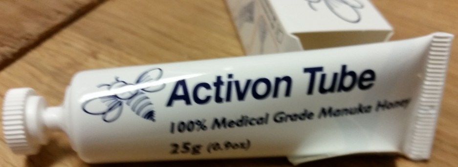
For oders of Manuka Honey click this website :http://www.highlandproductfinder.co.uk/

The study retrieved from:Manuka honey treatment of biofilms of Pseudomonas aeruginosa results in the
emergence of isolates with increased honey resistance
Annals of Clinical Microbiology and Antimicrobials 2014, 13:19 doi:10.1186/1476-0711-13-19
Aimee L Camplin (st10003013@outlook.uwic.ac.uk)
Sarah E Maddocks (smaddocks@cardiffmet.ac.uk)

Manuka honey-impregnated dressings in the treatment of neuropathic diabetic foot ulcers

Medicinal properties of honey have been known for millenniums and have been used for the treatment of a variety of pathological conditions (1). The healing properties of honey have also been known from long and recently there has been a resurgence of interest about the ability of this natural product to assist wound healing with numerous reports in the international bibliography (2) As a wound dressing, honey provides a moist environment with antimicrobial properties, has anti-inflammatory effects,reduces oedema and exudates, promotes angiogenesis and granulation tissue formation, induces wound contraction, stimulates collagen synthesis, facilitates debridement and accelerates wound epithelialisation (2–6). Honey efficacy in the healing of skin ulcers of different aetiologies has been documented in numerous studies (7).Antibacterial action of honeyhas been attributed to its hyperosmolarity, acidity or other properties that have not been fully elucidated (8). Hydrogen peroxide is produced upon dilution of honey (9) by the enzymatic activity of oxidases added in the nectar by bees (10), and it has been suggested to be the major antibacterial factor in at least some kind of honey. Apart from being an antiseptic H2O2 stimulates macrophage chemotaxis, induces Vascular Endothelial Growth Factor (VEGF) expression at the transcriptional level and consequently promotes angiogenesis and stimulates fibroblast proliferation while also possessing antioxidant action, protecting the local wound milieu from oxidative stress (11–13). Honey also exerts significant actions on the immune system, both innate and adaptive, stimulating cytokine production [TNFa, interleukin (IL)-1β and IL-6] by monocytes (13) and inducting B- and T-lymphocyte proliferation (14). The induction of proinflammatory cytokines by honey has also been reported to contribute to its antibacterial activity (13). The acidification of the alkaline environment of chronic non-healing ulcers by honey has also been proposed as another mechanism by which honey induces healing. Acidification inhibits protease activity, induces fibroblast proliferation and establishes an aerobic environment, all of which aid in the healing process (2).Nitric oxide (NO) is an important mediator in inflammation, cell proliferation and immune response and is actively implicated in wound healing (15,16). NO metabolites contained in honey (17) and induction of NO production by honey in different body fluids (18) constitute another mechanism by which honey induces wound healing, given the antimicrobial and immunoregulatory actions of NO. Manuka honey (MH) is a natural, monofloral honey produced from bees feeding on manuka (Leptospermum scoparium) plant which is endemic in parts of Australia and New Zealand. MH has been reported to exhibit antibacterial activity against a broad spectrum of bacteria including Staphylococcus aureus [including Methicillin-Resistant Staphylococcus aureus (MRSA)], Pseudomonas aeruginosa and vancomycinsensitive and vancomycin-resistant enterococci (19–22). MH has been found to arrest cell-cycle progression and prevent cell division of S. aureus (23) and to induce cell disruption and lysis of P. aeruginosa cells (24). Methylglyoxal has been identified as the active antibacterial component of MH (25,26). Diabetic foot ulcers are reported to occur in 15% of patients (with different frequencies between type I and II diabetic patients) with diabetes and to antedate 84% of all diabetes-related amputations (27,28). Peripheral neuropathy leading to unperceived trauma seems to be the major cause of diabetic foot ulcers with 45–60% of ulcers to be considered merely neuropathic and 45% of mixed, neuropathic and ischaemic aetiology (29,30). Lower extremity ulcers represent one of the most common complications of diabetes and a leading cause for hospitalisation of diabetic patients (31). Neuropathy, deformity, high plantar pressure, poor glycaemic control, long duration of diabetes, peripheral arterial disease, and male gender all are risk factors for lower extremity
ulceration (27,31,32). Treatment of lower extremity ulcers imposes a huge burden on health care systems worldwide with at least 33% of all costs to treat diabetes complications to be spent for the treatment of ulcers (33). MH has been reported to be effective in the treatment of leg ulcers of diverse aetiology (2,34,35) and is considered as honey with high antibacterial properties.
For orders you may visit this website: http://www.highlandproductfinder.co.uk/

The study is retrieved from :Manuka honey-impregnated dressings in the treatment of
neuropathic diabetic foot ulcers (Alexandros V. Kamaratos1, Konstantinos N. Tzirogiannis1, Stella A. Iraklianou1,
Georgios I. Panoutsopoulos2, Ilias E. Kanellos1 & Andreas I. Melidonis1, 2012)